Technologies
Histopathology & in-situ Hybridisation
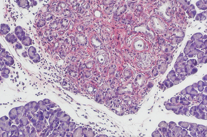
Histology is the study of the anatomy of cells and tissues of plants and animals using microscopy. It is commonly studied using a light microscope or electron microscope, the specimen having been sectioned, stained, and mounted on a microscope slide. Histological studies may be conducted using tissue culture, where live animal cells are isolated and maintained in an artificial environment for various research projects. The ability to visualize or differentially identify microscopic structures is frequently enhanced through the use of staining. Histology is an essential tool of biology and medicine.
Histopathology, the microscopic study of diseased tissue, is an important tool in anatomical pathology, since accurate diagnosis of cancer and other diseases usually requires histopathological examination of samples. (https://en.wikipedia.org/wiki/Histology)
Areas of Technology Expertise:
Tissue Processing
- Sectioning (to include microtomy, cryotomy, vibratome and resin sectioning)
- Paraffin processing/embedding
- Tissue freezing
- Histochemical staining (H&E and special stains)
- Tissue Microarrays (TMA)
Laser Capture Microdissection (LCM)
Immunohistochemistry (IHC) including immunofluorescence and multiplexing
- Immunohistochemical staining – localization of biomarkers and differentially expressed proteins in different parts of a biological tissue.
- Immunostaining with antibodies conjugated to an enzyme, such as peroxidase or to a fluorophore (Immunofluorescence)
- TUNEL staining
In situ Hybridisation (ISH) including FISH, miRNA ISH, RNAscope, BaseScope and In Situ Mutation Detection (ISMD)
Image Processing
-
-
- Slide Scanning (Brightfield & Fluorescence)
- Image analysis (e.g. Indica Labs HALO)
- Image sharing (e.g. Phillips Xplore)
- Mouse and Human Pathology
-
Facilities/cores in Cambridge which can provide skills, knowledge or access to equipment:
CRUK CI – www.cruk.cam.ac.uk/core-facilities/histopathology-core
Metabolic Research Laboratories (MRL), University of Cambridge, Wellcome Trust-MRC Institute of Metabolic Science – www.mrl.ims.cam.ac.uk/research/core-facilities/histology-core/
Addenbrooke’s Tissue Bank – https://www.cuh.nhs.uk/tissue-bank
Wellcome-MRC Stem Cell Institute – https://www.stemcells.cam.ac.uk/research/facilities/histology
Category Histology
Date 24 February 2017
Technology MRI, etc.